Figure 3.
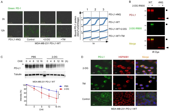
2-DG decreases PD-L1 translocation and stabilization. A. Left, The quantitative binding of PD-1 Fc protein on PD-L1-overexpressing MDA-MB-231 cells was assessed at the indicated times. Cells were treated with 10 mmol/L 2-DG, 1 µgml-1 tunicamycin (TM), or 10 µmol/L olaparib. Right, Images of PD1 Fc protein on PD-L1-overexpressing MDA-MB-231 cells from 0 to 72 hours. B. Western blot analysis of PD-L1 protein expression in PD-L1-overexpressing MDA-MB-231 cells and MDA-MB-231-4NQ cells treated with 2 mmol/L 2-DG IR800. C. Western blot analysis of PD-L1 protein expression in PD-L1-overexpressing MDA-MB-231 cells. Cells were treated with 20 mM cycloheximide (CHX) with or without 2 mmol/L 2-DG at the indicated times. The intensity of PD-L1 protein expression was quantified using a densitometer. *P<0.05. D. Confocal microscopy image showing HSP90B1 and PD-L1 expression in PD-L1-overexpressing MDA-MB-231 cells after treatment with 2-DG. Scale bar, 20 mm.

