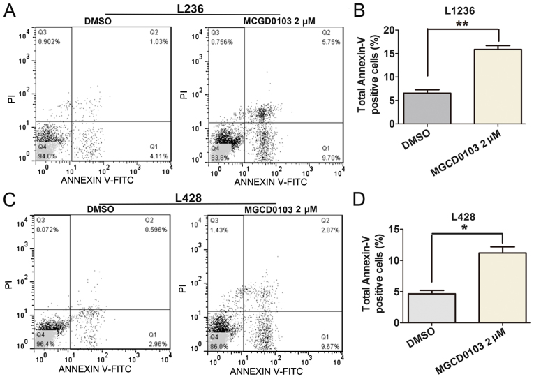
Figure 4.
Apoptotic rates were determined by flow cytometry with Annexin-V/PI double-staining. In the MGCD0103 2 µM group, cells were treated with MGCD0103 at a concentration of 2 µM, with the DMSO group considered the control group; cells were treated for 24 h. Total percentages of Annexin-V-positive cells in DMSO and MGCD0103 groups were measured in (A and B) L1236 and (C and D) L428 cells. The percentage of Annexin-V-positive cells in the DMSO groups was lower than that of the MGCD0103 group, in both cell lines. Data are presented as the mean + the standard error of the mean. *P<0.05 and **P<0.01. PI, propidium iodide; DMSO, dimethyl sulfoxide; FITC, fluorescein isothiocyanate.

