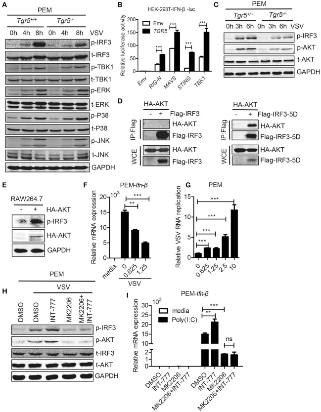
Figure 6.
TGR5 amplifies IFN-I signaling via AKT-mediated IRF3 activation. (A) Immunoblot analysis of phosphorylated (p-) or total (t-) proteins in lysates of Tgr5+/+- and Tgr5−/−-PEMs infected with VSV (1 MOI) for the indicated times. (B) TGR5 was co-transfected with RIG-I (N) (RIG-N), MAVS, STING, TBK1, or empty vectors, together with an IFN-β luciferase reporter, into HEK-293T cells for 28 h. IFN-β luciferase activity was detected and normalized to Renilla luciferase activity. (C) Immunoblot analysis of phosphorylated (p-) AKT or total (t-) AKT in lysates of Tgr5+/+- and Tgr5−/−-PEMs infected with VSV (1 MOI) for the indicated times. (D) HEK-293T cells were transfected with plasmids encoding HA-AKT and Flag-IRF3 or Flag-IRF3-5D for 28 h. The cell lysate supernatants were immunoprecipitated using M2 beads, and then immunoblotted with antibodies to HA or Flag tags. WCE, whole-cell extracts. (E) Immunoblot analysis of phosphorylated (p-) IRF3 and HA-AKT in lysates of RAW 264.7 cells transfected with plasmids encoding HA-AKT for 28 h. (F) Q-PCR analysis of Ifn-β expression in PEMs pretreated with MK 2206 (an inhibitor of AKT) at the indicated dose (μM) for 1 h and then infected with VSV (1 MOI) for 8 h. (G) Q-PCR analysis of VSV RNA replicates in PEMs pretreated with MK 2206 at the indicated dose (μM) for 1 h and then infected with VSV (1 MOI) for 8 h. (H) Immunoblot analysis of phosphorylated (p-) IRF3 and (p-) AKT in lysates of PEMs pretreated with MK 2206 (3 μM) or INT-777 (500 μM) for 1 h, and then infected with VSV (1 MOI) for 8 h. (I) Q-PCR analysis of Ifn-β expression in PEMs pretreated with MK 2206 (3 μM) or INT-777 (500 μM) for 1 h, and then stimulated with poly (I:C) (10 μg/ml) for 2 h. GAPDH was used as an internal control for Q-PCR. The data are shown as the mean ± SD. ns, not significant; **P < 0.01; ***P < 0.001. All experiments were performed three times with similar results.

