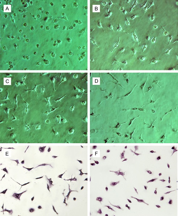
Figure 1.

Purified cultures of microglia (phase contrast microscope, ×250). (A) Microglia were round and irregular at day 1; (B) A few microglia extended protuberances at day 2; (C, D) Cell bodies were elongated, and asymmetrical branches were visible at day 4 (C) and day 5 (D); (E) On day 5, microglia were positive for CD11b/c; (F) On day 5, microglia were positive for NF-кB p65 in the cytoplasm.
