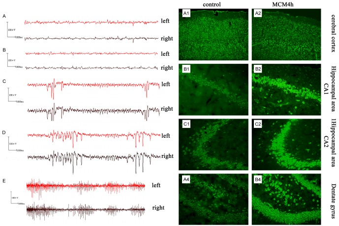
Figure 5.
EEG recording and glutamate (Glu) expression in brain tissue after intraventricular injection with microglia-conditioned medium (MCM). (A-E) EEG showed epileptiform waveforms after MCM injection. (A) EEG from the control group. (B-D) EEG at 45 min after intraventricular injection in the MCM0 (B), MCM2 (C) or MCM4 (D) group. No significant difference in potential waveform or amplitude between the control group and MCM0 groups was detected. Epileptiform waveforms (e.g., spike wave, sharp wave and spike-slow wave) were seen in the MCM2 and MCM4 groups. (E) EEG at 1 h after intraventricular injection in the MCM4 group displayed enhanced amplitude and sharp waves. (A, B) Expression of Glu in rat cerebral cortex and hippocampus, determined by immunofluorescence. Glutamate content in was increased to different degrees in all areas compared with the control group 45 min after injection with MCM4. (A1-4) control group; (B1-4) MCM4 group. (B1) Cerebral cortex, ×100; (B2) Hippocampal CA1 region, ×200; (B3) Hippocampal CA3 region, ×200; (B4) Dentate gyrus, ×200.

