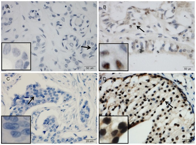
Figure 1.
Representative micrographs showing (A and C) negative and (B and D) positive immunohistochemical staining of poly(ADP-ribose) polymerase-3 in (A and B) tumor-adjacent tissues and (C and D) breast cancer tissues. Arrows indicate the magnified regions in the insert. Magnification, ×400. Scale bar, 50 µm.

