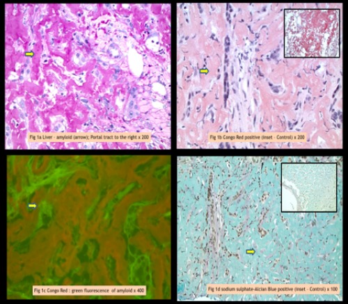
Figure 1.

A) Extensive amyloid within liver parenchyma, appearing as amorphous eosinophilic waxy deposits (arrow). Adjacent portal tract is clear H&E stain. B) Brick red staining (arrow) of amyloid deposits using Congo red (Inset shows positive control); C) Amyloid deposits emitting green fluorescence with Congo red stain (arrow); D) Amyloid deposits staining bluish-green (arrow) with Sulfated Alcian Blue (SAB) stain (Inset shows positive control).
