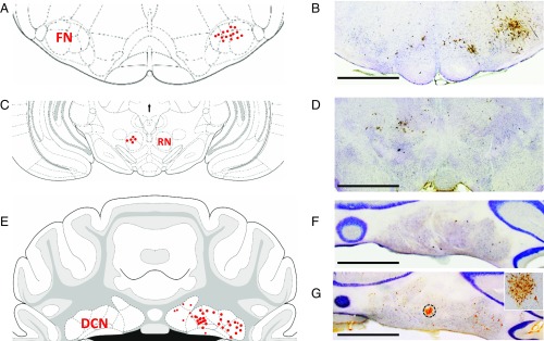
Fig. 2.
Identification of DCN projection neurons by the transsynaptic neuronal tracer PRV-152. (A, C, and E) The distribution of PRV-labeled neurons in FN (A), RN (C), and DCN (E). (B, D, F, and G) Representative PRV-labeled neurons in the FN (B), RN (D), and DCN (F and G) which were identified by anti-PRV immunolabeling. Atlas images were modified from ref. 79. All light microscope images were taken with a 1.25× microscope objective. (Scale bars: 500 µm.) The Inset in G was taken with microscope with a 10× objective from the DCN area indicated by dashed circle. Note that PRV-152 was injected into orbicularis oculi muscle of the upper eyelid, taken up by axon terminals of FN neurons, retrogradely transported to the RN, and reached the DCN after 3 d of PRV inoculation. Extensive PRV-infected DCN neurons were observed after 3.5 d of PRV inoculation.

