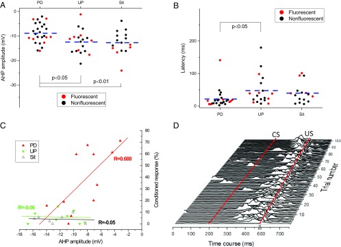
Fig. 4.
Delay EBC produces learning-related changes in membrane properties of evoked DCN AP. (A–C) Learning-related changes in the evoked AP as a result of delay EBC were characterized by alterations in AHP amplitude (A), latency for evoked AP (B), and a positive linear association between AHP amplitude and percent CRs (CRs%) at the fourth session (C). Note that each point in the A and B represents the result from a single neuron (red solid circles for fluorescent neurons and black solid circles for nonfluorescent neurons), and the dashed line illustrates the average for each group. Each point in C represents the result from a single animal. (D) Representative eyelid EMG activity from a rat given paired conditioning during the last training session. Note that the rat had 71% CRs and a reduced AHP amplitude of −3.17 mV. Dashed lines indicate the onset times of the CS and US. A blanking circuit in operation during the US (the break in the x axis) prevented the shock from saturating the EMG amplifier.

