Fig. 5.
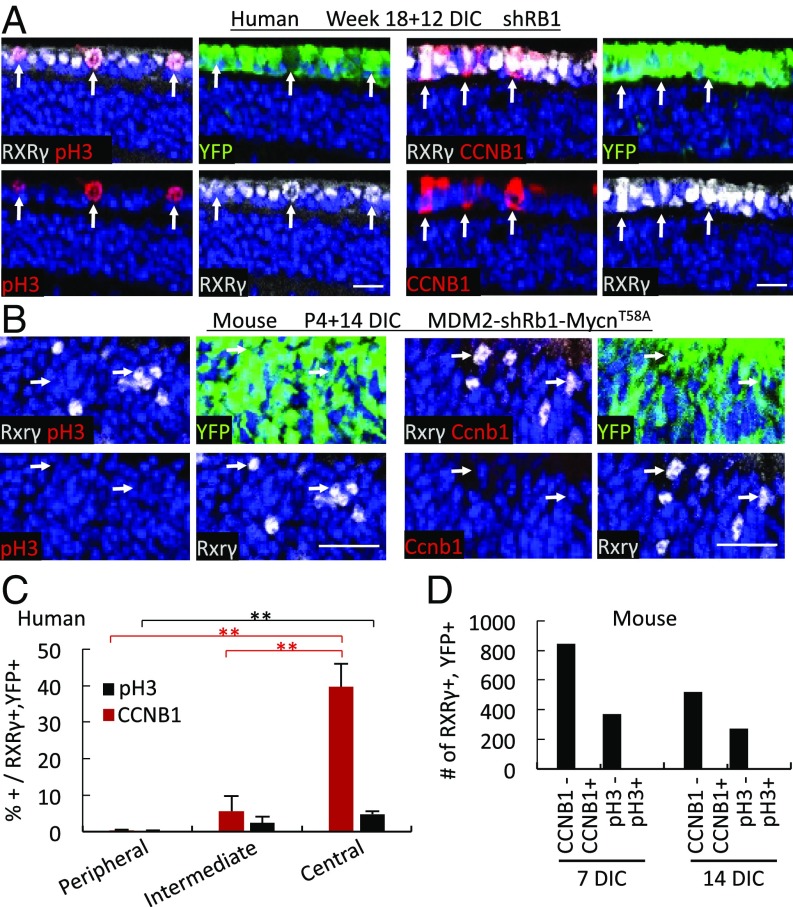
Rb-depleted human but not mouse cone precursors express G2 and M-phase markers. (A and B) Cyclin B1 (CCNB1) and phospho-histone H3-Ser10 (pH3) (red) in RXRγ+ (white) cone precursors of 18 wk human retinae at 12 d after RB KD (A) but not in Rxrγ+ (white) cone precursors of P4 RGP-MDM2 mouse retinae at 14 d after Rb KD and Mycn overexpression (B). (Scale bars, 20 μm.) (C) The percentage of CCNB1+ or pH3+ cells among RXRγ+,YFP+ cone precursors in human retina sections adjacent to those stained for Ki67 and EdU in Fig. 1F. (D) The number of RXRγ+,YFP+ cells analyzed for CCNB1 or pH3 in RGP-MDM2 retinae cotransduced with shRb1 and MycnT58A at 7 and 14 DIC. Error bars indicate SD. Significance was assessed by t test (**P < 0.001).

