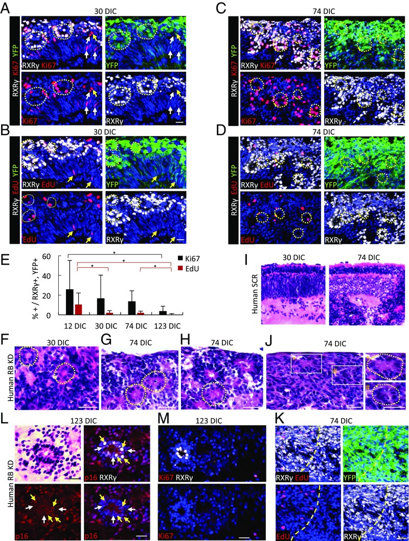
Fig. 6.
Cone precursor proliferation and retinoma genesis in RB-depleted human but not mouse retinae. (A–D) Ki67 expression (A and C) and EdU incorporation (B and D) (red) at 30 DIC (A and B) and 74 DIC (C and D) after transduction of week 18 fetal retinae. Dashed circles identify rosette-like arrangements of RXRγ+ nuclei. Arrows indicate proliferating transduced (yellow arrows) or nontransduced (white arrows) noncone (RXRγ−) cells likely representing progenitors or glia. (E) Quantitation of Ki67 expression and EdU incorporation in RXRγ+ cone precursors in week 18 retinae at 12–123 DIC. Data at each age are from central, intermediate, and peripheral regions. Error bars indicate SD. Significance was assessed by ANOVA with Tukey’s HSD post hoc test (*P < 0.05). (F–J) H&E staining of week 18 retinae at the indicated DIC after transduction with shRB1 (F–H and J) or shSCR (I). Dashed circles identify Flexner–Wintersteiner rosettes (F–H) or fleurettes (J, Right). (Left) Enlarged views of boxed regions. (K) Predominance of RXRγ+,YFP+ cells in the retinoma-like region shown in J. The dashed yellow line separates proliferating (upper left) and nonproliferating (lower right) regions. (L) H&E staining (Upper Left) performed after p16INK4A immunostaining and imaging of RB-depleted retina at 123 DIC, showing a central fleurette with predominantly cytoplasmic p16 in RXRγlo cells (white arrows) and nuclear p16 in RXRγhi cells (yellow arrows). (M) A fleurette composed of RXRγ+,Ki67− cells at 123 DIC. (Scale bars, 20 µm.)

