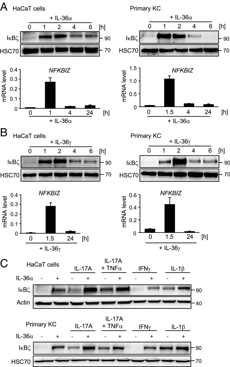
Fig. 1.
IL-36 induces IκBζ expression in KCs. (A and B) HaCaT cells (Left) or human primary KCs (Right) were treated with 100 ng/mL IL-36α (amino acids 6–158) (A) or 100 ng/mL IL-36γ (amino acids 18–169) (B) for the indicated times. IκBζ protein was analyzed by Western blotting. Relative mRNA levels of NFKBIZ were measured in parallel and normalized to the reference RPL37A. (C) HaCaT cells (Upper) and primary KCs (Lower) were treated for 2 h with 100 ng/mL IL-36α alone or in combination with 100 ng/mL IL-17A, 10 ng/mL TNFα, 100 ng/mL IFNγ, or 100 ng/mL IL-1β. IκBζ was detected by Western blotting. HSC70 or β-actin served as loading controls.

