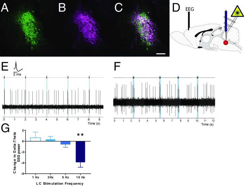
Fig. 1.
Selective optogenetic control of LC-NE neurons at subarousal levels. LC neurons identified with TH staining (A, green) were transduced to selectively express ChR2-mCherry (B, magenta) at high levels (C, merge). (Scale bar: 100 μm.) (D) Schematic of glass pipette optrode and PFC EEG recording configurations. (E) Pulses of blue light elicited reliable short-latency single-action potentials from LC neurons in vivo (Inset, LC unit waveform) at 1 Hz up to 15 Hz. (F) Short trains of 15-Hz phasic light pulses evoked phasic excitation of LC neurons, followed by a postactivation inhibition pattern comparable to innate phasic LC responses to salient stimuli. (G) Increases in tonic LC stimulation dose-dependently increased arousal, as seen by a reduction in δ-dominant EEG recordings at frequencies >5 Hz. **P < 0.01 relative to baseline arousal.

