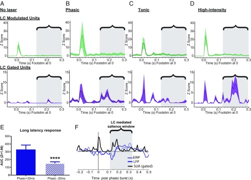
Fig. 4.
Phasic, but not tonic, LC activity generates salience-like features in S1. (A) Response ± 95% confidence interval (CI) to low-intensity hind-paw stimulation without LC photoactivation for LC-modulated neurons (Top, green) and LC-gated neurons (Bottom, purple) averaged across 50 trials and locked to foot stimulus (Footstim) at time 0. The bracketed region with gray boxes throughout highlights the phasic LC-mediated salience window for coordinating responses across cortical targets within which long-latency responses were identified. (B) Response ± 95% CI to low-intensity hind-paw stimulation with time-locked phasic LC photoactivation for LC-modulated neurons (Top, green) and LC-gated neurons (Bottom, purple) averaged across 50 trials and locked to Footstim at time 0. (C) Response ± 95% CI to low-intensity hind-paw stimulation with time-locked tonic LC photoactivation for LC-modulated neurons (Top, green) and LC-gated neurons (Bottom, purple) averaged across 50 trials and locked to Footstim at time 0. (D) Response ± 95% CI to high-intensity hind-paw stimulation without LC photoactivation for LC-modulated neurons (Top, green) and LC-gated neurons (Bottom, purple) averaged across 50 trials and locked to Footstim at time 0. Note the prominent long-latency response found in B and D with phasic LC stimulation around 200 ms after the hind-paw stimulus. (E) Comparison of the long-latency response seen from LC-gated neurons in B after a temporal shift in phasic LC photoactivation from +20 ms to −20 ms before Footstim. The magnitude of the long-latency response was markedly reduced by this temporal offset. ****P < 0.0001. (F) Temporal overlay of ERP response from mPFC, LFP response from S1, and single-unit activity (SUA) response from LC-gated S1 neurons aligned to the onset of phasic LC bursting at time 0.

