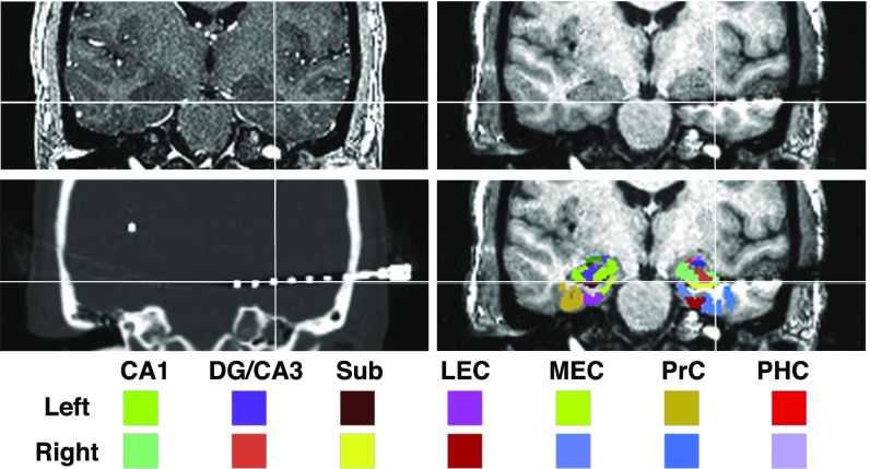
Fig. 3.
Examples of MRI, CT scan, and template for a single subject. Electrodes were localized in each subject using coregistered preimplantation (Upper Left), postimplantation MRI (Upper Right), and CT (Lower Left) scans. A high-resolution template labeled with medial temporal lobe (MTL) subregions was aligned to each subject’s preimplantation scan to guide electrode localization. Regions of interest in the MTL included the CA1, DG/CA3, subiculum (Sub), lateral and medial entorhinal cortex (LEC, MEC), and the PRC and PHC cortices. The template also provided labels for amygdala nuclei, which can be seen in the figure but were not included in the analysis.

