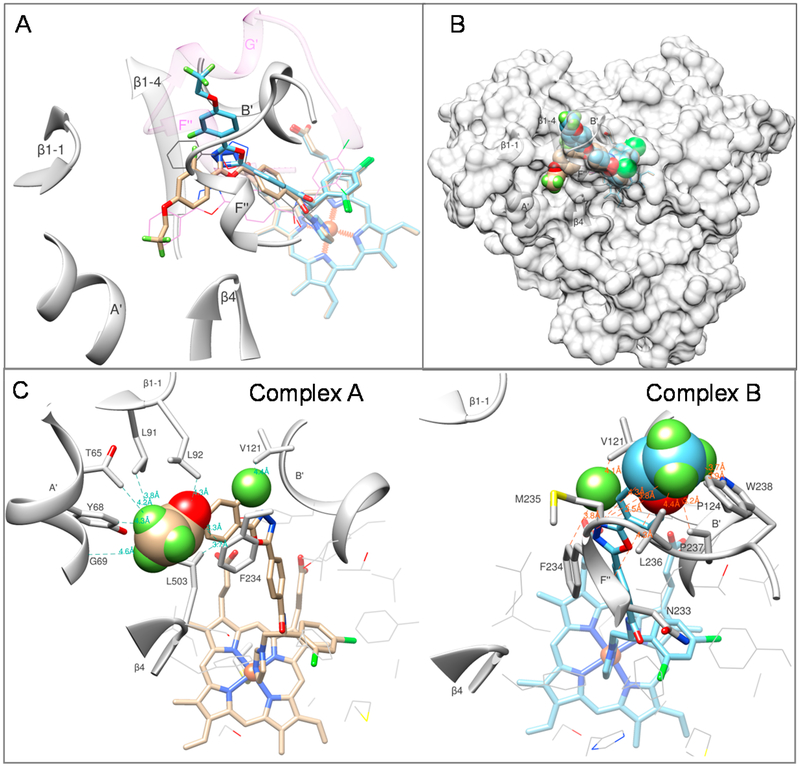
Figure 3.
LFV (compound 3) adopts two alternative conformations in the active site of A.fumigatus CYP51B. (A) Substrate entry area. The carbon atoms in the superimposed structures of complexes A and B are tan and blue, respectively. The corresponding orientation of VNI in the structures of A.fumigatus CYP51B (PDB ID 4uyl, gray lines) and Trypanosoma brucei CYP51 (PDB ID 3gw9, purple lines) are shown for comparison. The A. fumigatus CYP51 substrate entry-defining secondary structural elements are presented as gray ribbon and labeled. The semitransparent ribbon of the (longer) F/G loop area in the protozoan CYP51 (3gw9) is purple, and the F′′ helix and the protozoan-specific G′ helix are labeled. (B) Overall view in semitransparent surface representation. (C) New ligand/enzyme interactions formed as a result of the VNI modification. Atoms added to the VNI structure are presented as spheres. The amino acid residues of A. fumigatus CYP51B involved in the additional interactions are shown as stick models and labeled, and selected distances are displayed.

