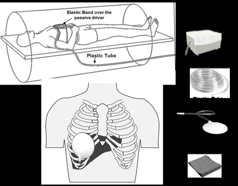
Fig.1.

MRE of liver set up. Note that active driver is placed outside the scanner and mechanical waves are conducted into the passive driver via a long plastic tube. The passive driver is placed at the level of xiphisternum and in right mid clavicular line as shown.
