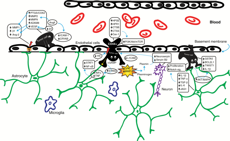
Fig. 1.
Schematic representation of the stages of the formation of cerebral metastasis. From left to right, the processes of vascular adhesion, transgression through the BBB, extravasation, interaction with the brain microenvironment of the tumor cells and involved molecules are presented. MMP, matrix metalloproteinase; β1, beta 1 integrin subunit; PTGS2/COX-2, prostaglandin-endoperoxide synthase 2/cyclooxygenase-2; ADAM8, a disintegrin and metalloproteinase domain-containing protein 8; β4, beta 4 integrin subunit; CRYAB, αβ-crystallin; INFα, interferon alpha; STING, stimulator of interferon genes; L1CAM, L1 cell adhesion molecule; JAG1, jagged 1; GSTA5, glutathione S-transferase alpha 5; BCL2L1, BCL2 like 1; TWIST1, twist family BHLH transcription factor 1.

