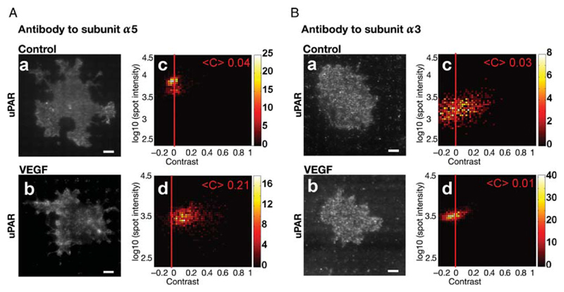
Figure 4.
VEGF-induced interaction of uPAR with the fibronectin receptor integrin-α5β1. HUVECs were grown on microchips patterned with antibodies against α5- (A) or α3-integrins (B). After stimulation with VEGF165 (50 ng/mL) for 60 min, cells were fixed and immunostained with a fluorescent monoclonal antibody against uPAR. Representative TIRF images visualize VEGF165-induced uPAR redistribution to mimic the pattern of the antibody to integrin subunit α5 (cf. A.a and A.b), which is absent on surfaces coated with an antibody to integrin subunit α3 (cf. B.a and B.b); scale bar = 6 μm. Colour density plots (A.c, A.d; B.c, B.d) summarize the data from 100 to 120 cells.

