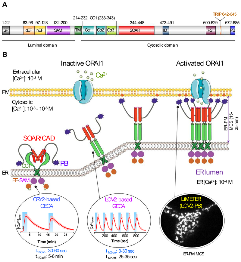
Figure 1 ∣. Engineering STIM1 to convert SOCE at ER-PM membrane contact sites (MCSs) into light-operated Ca2+ entry.
(A) Schematics of the domain architecture of human STIM1. The luminal domain of STIM1 contains a signal peptide (SP), a hidden non-Ca2+ binding EF-hand motif (hEF), a canonical Ca2+ binding EF-hand motif (cEF), and a sterile alpha motif domain (SAM). The cytoplasmic domain comprises a putative coiled-coil region (CC1), a minimal STIM1 ORAI-activating region (SOAR or CAD or OASF), an inactivation domain (ID), a proline/serine-rich region (PS), an EB1-binding sequence (TRIP), and a C-terminal polybasic tail (PB).
(B) Cartoon illustration of the dynamic coupling between STIM1 and ORAI1 during SOCE activation at ER-PM apposition, a specialized membrane contact site that is separated by a distance of approximately 25-35 nm without membrane fusion. The major steps in this tentative model include: (i) Ca2+ depletion induces oligomerization of the luminal EF-SAM domain to initiate STIM1 activation; (ii) the luminal signal is transduced toward the cytoplasmic side to overcome STIM1 autoinhibition mediated by the intramolecular CC1-SOAR interaction, thereby triggering a conformational switch to expose SOAR/CAD/OASF and the C-terminal PB domain; (iii) activated STIM1 undergoes further oligomerization and its subsequent migration toward the PM is facilitated by the association between the positively charged PB domain and PM-embedded, negatively-charged phosphoinositides. SOAR/CAD/OASF is responsible for directly engaging and activating ORAI channels to mediate Ca2+ flux from the extracellular space into the cytosol. When coupled to plant-derived photosensory domains, such as cryptochrome 2 (CRY2) and light-oxygen-voltage domain 2 (LOV2), these critical steps can be individually mimicked with engineered Opto-CRAC constructs (CRY2-STIM1 chimeras or LOV2-STIM1 chimeras) and ER-tethered LOV2-PB proteins (designated LiMETER for light-inducible membrane-tethered peripheral ER). Representative Ca2+ signals generated by Opto-CRAC variants following repeated light-dark cycles are shown with the circles, along with a typical image showing the light-induced assembly of ER-PM MCSs in HeLa cells revealed by total internal reflection fluorescence (TIRF) microscopy. These constructs are available at Addgene (accession ID: #101245, #101246, #113933 and #113934).

