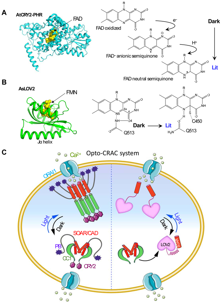
Figure 2 ∣. Opto-CRAC for optical control of Ca2+ influx in mammalian cells.
(A-B) The 3D structures and simplified photocycle reactions of AtCRY2-PHR (modeled from CRY1; PDB entry: 1U3D; A) and AsLOV2 (PDB entry: 2V0W; B). FAD, flavin adenine dinucleotide; FMN: flavin mononucleotide.
(C) The left semi-circle depicts an Opto-CRAC construct made of a CRY2-STIM1 fusion. Upon light illumination, CRY2 undergoes oligomerization, mimicking Ca2+-depletion induced EF-SAM multimerization, to trigger STIM1 activation with subsequent opening of ORAI Ca2+ channels situated in the plasma membrane. The right semi-circle illustrates the engineering strategy of an alternative form of Opto-CRAC, in which the CC1-SOAR interaction-mediated intramolecular autoinhibition is recapitulated by LOV2-SOAR fusion in the dark. Following photostimulation, LOV2 undergoes rapid conformational changes to expose the C-terminal effector domain, thereby restoring the potent ORAI-activating activity of SOAR to elicit Ca2+ influx.

