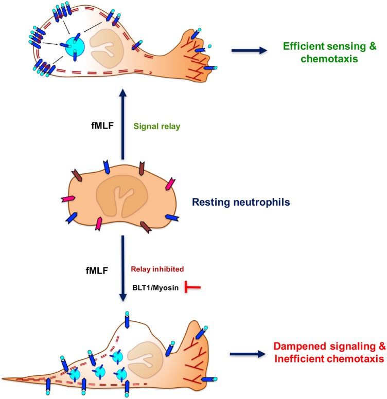
Fig. 7.
Schematic representation of PMNs internalizing FPR1 from the trailing edge of a neutrophil and the contribution of the BLT1-myosin axis in restricting the extent of FPR1 internalization to promote the directional migration toward fMLF. Elongated chevrons with different colors in the resting neutrophils represent ligand-free GPCRs. The blue chevrons with aqua circles represent fMLF-bound FPR1 in stimulated neutrophils. Double dashed maroon lines represent F-actin networks at the back of the cell. Maroon-colored ‘Y’ shapes represents dynamic and branched F-actin at the protrusive front of a stimulated neutrophil.

