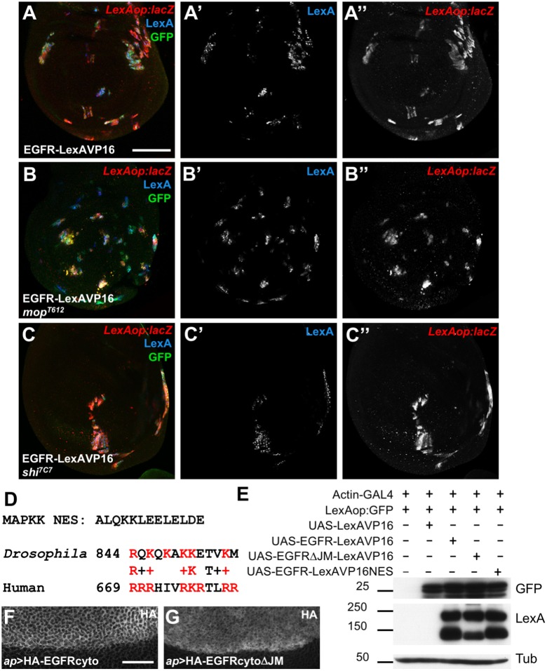
Fig. 2.
Overexpressed EGFR–LexAVP16 can bypass normal trafficking. (A–C) Wing discs in which UAS-EGFR-LexAVP16 is expressed in clones of cells that are wild type (A), mop mutant (B) or shi mutant (C). Clones are marked by anti-GFP antibody staining (green) and anti-LexA antibody staining (A′–C′, blue). Anti-β-gal antibody staining reflects LexAop:lacZ expression (A″–C″, red), which is not reduced by the blocking of EGFR cleavage that occurs in mop cells or of endocytosis that occurs in shi cells. (D) Sequence of the NES from MAPKK and an alignment of the amino acids immediately following the transmembrane domains of Drosophila and human EGFR. Basic residues are in red. (E) Western blots of S2R+ cell lysates transfected with the indicated constructs. Deleting the juxtamembrane region reduces EGFR–LexAVP16 cleavage, but neither this deletion nor adding an NES reduces reporter expression. (F,G) Portions of wing discs spanning the dorsal-ventral boundary in which UAS-HA-EGFRcyto (F) or UAS-HA-EGFRcytoΔJM (G) is expressed in the dorsal (upper) compartment with ap-GAL4. Anti-HA antibody staining shows cytoplasmic localization. Scale bars: 50 μm (A), 20 μm (F).

