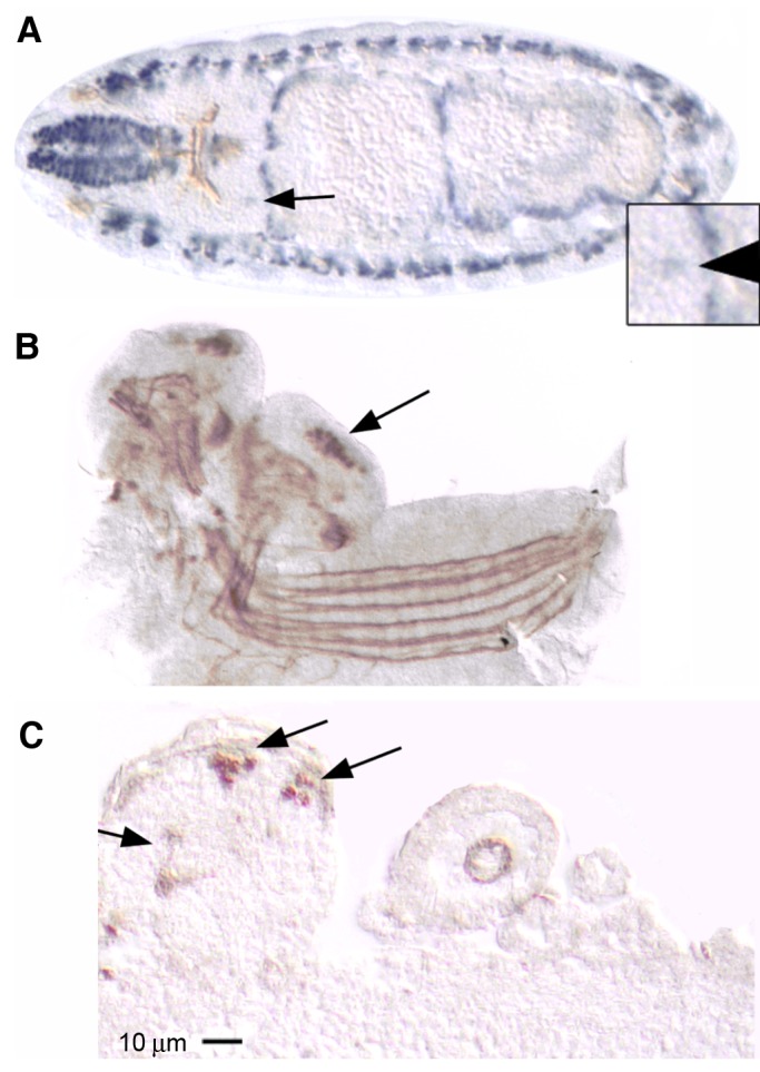
Fig. 5.

MEF2 is expressed in embryonic mushroom bodies. (A) A horizontal section through a stage 15 wild-type embryo embedded in plastic and immunostained for MEF2 (alkaline phosphatase-coupled secondary antibody, blue) and FASII (horseradish peroxidase-coupled secondary antibody, brown). MEF2 expression is abundant in somatic and visceral muscle cell nuclei and is also visible in bilaterally symmetrical cells of the dorso-posterior brain where MB neurons are localized (arrow and magnified in inset). FASII labels axon tracts throughout the developing nervous system whereas MEF2 is localized to cell nuclei. (B) A wholemount of the central nervous system dissected from a late stage 17 wild-type embryo and immunolabeled for MEF2 and FASII (both detected with horseradish peroxidase-coupled secondary antibody substrate, brown). MEF2 and FASII immunoreactivity is distinguished by the respective localization to nuclei and axons. MEF2 expression is highly enriched in the MB nuclei (arrow). The brain is slightly turned so that both hemispheres are equally visible. (C) A sagittal paraffin section through a late stage 17 embryo that is heterozygous for the mef226-49 mutation in which MEF2 is mislocalized to the cytoplasm. MEF2 immunoreactivity is apparent in the pedunculus and vertical lobe (arrow) and MB nuclei (arrows in dorso-posterior brain). Anterior is to the left in A–C. At least three embryos showed similar results for each experiment.
