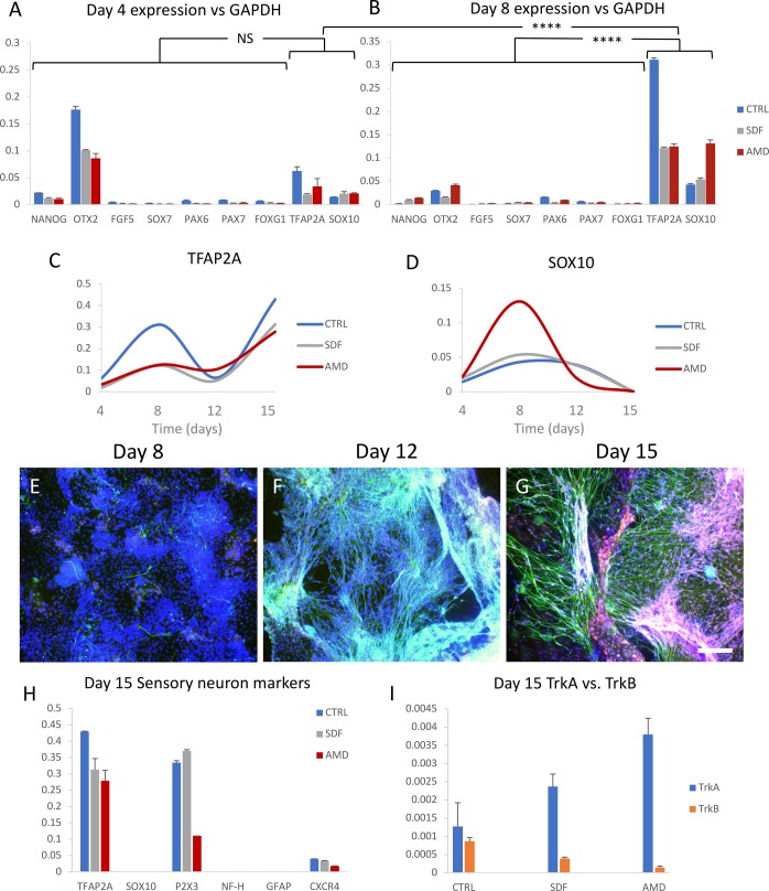
Fig. 1.
Directed differentiation of hiPSCs to sensory neurons under CXCR4 stimulation or inhibition. Differentiation cultures from four time-points (4, 8, 12 and 15 days) were cultured, each with three groups [control, inhibitor (AMD3100) and agonist (SDF1)] with six experimental replications each (72 cultures total). (A,B) Transcription level of various markers relative to GAPDH in the three differentiation groups on days 4 and 8 shows the transition to neural crest cells (TFAP2A/SOX10). Grouping neural crest markers shows their expression to be not significantly (NS) higher than non-neural crest markers on day 4, but highly significant (****P<0.0001) by day 8. (C,D) expression of TFAP2A and SOX10 in the three groups relative to GAPDH over four time-points (day 4, 8, 12 and 15). (E–G) Sensory neuron differentiation progression of the control group with nuclei in blue, neurites (beta-III-tubulin) in green, and TrkA in red (sensory neurons, G only) on days 8, 12 and 15. (H) Transcription levels of markers relevant to sensory neuron differentiation and subtype specification relative to GAPDH on day 15. (I) Transcription levels of TrkA and TrkB in the three groups relative to GAPDH on day 15. qPCR: single cDNA pool from two replications, error bars: standard deviation of qPCR replications. Scale bar: 150 µm.

