
Fig. 5.
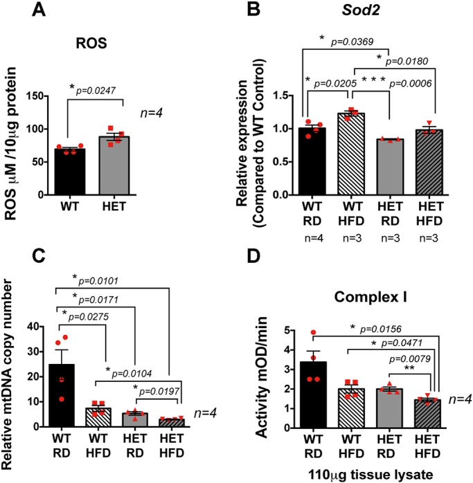
Reduced antioxidant capacity in RPE/retina of Pgc-1α+/− mice, and decreased mitochondrial activity in RPE/retina of Pgc-1α+/− mice fed HFD. (A) ROS measurement in the RPE/retina extract of WT and Pgc-1α+/− mice showing increased ROS levels in the Pgc-1α+/− mice as compared with WT. (B) Reduced antioxidant capacity in the RPE/retina of Pgc-1α+/− mice, as compared with WT, shown by reduced Sod2 expression and inability to induce Sod2 expression under stress conditions such as HFD. (C) mtDNA copy number measured by qPCR showing reduced mtDNA induced by HFD. Pgc-1α repression combined with HFD further reduced the mtDNA copy number. (D) Pgc-1α repression and HFD significantly reduced mitochondrial complex I activity.