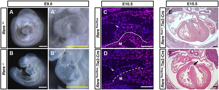
Fig. 3.
Ablation of Rere in the endocardium leads to the development of VSDs. To test the ability of the Rere flox allele to generate a null allele after exposure to Cre, we generated a line of Rere+/− mice by crossing Rereflox/flox mice with mice carrying an Hprt-Cre allele (Tang et al., 2002). (A,A′) As expected, E9.0 Rere+/− embryos had normal cardiac looping. (B,B′) E9.0 Rere−/− embryos showed failure of cardiac looping, as previously documented in embryos that are homozygous for the om null allele (Zoltewicz et al., 2004). Scale bars: 0.5 mm. (C,D) Sagittal cardiac sections were prepared from E10.5 Rereflox/flox and Tie2-Cre; Rereflox/flox embryos and stained with anti-RERE antibodies. Rereflox/flox;Tie2-Cre embryos showed a tissue-specific decrease in RERE immunoreactivity in the endocardium and in the mesenchymal cells of the AV endocardial cushions when compared with Rereflox/flox littermate control embryos. Arrows indicate the endocardium; dashed lines mark the boundary of the myocardium and the AV endocardial cushions. M, myocardium. Scale bars: 100 µm. Representative images are shown from the analysis of at least nine sections obtained from each of three embryos. (E) E15.5 Rereflox/+;Tie2-Cre embryos have normal ventricular septums. (F) Approximately 33% (2/6) of Rereflox/flox;Tie2-Cre embryos at E15.5 have VSDs (black arrow). Scale bars: 200 µm.

