Fig. 5.
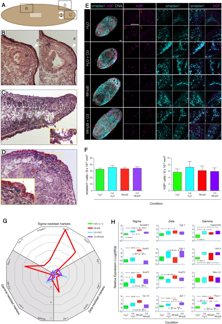
Smed-MmpB KD promotes tissue invasion and tumor formation. (A) Localization of the lesions shown in B and C. (B) Sagittal section of the dorsal posterior region stained with H&E. The large outgrowth (white arrowheads) contained ectopic structures such as photoreceptors (pr), neural tissue (nt) and a pharyngeal pocket (f), resembling a teratoma (d, dorsal). (C) Sagittal section of the median anterior region stained with H&E. The lesions observed were characterized by the displacement of gastrointestinal tissue that ruptured the epidermal layer [magnification, 40× (inset, 100×)]. (D) Frontal section of the anterior region stained with H&E. Epidermal blisters accumulated in the region anterior to the pharynx and were characterized by the presence of small cells that crossed the basal membrane, without altering the epidermal layer [magnification, 40× (inset, 100×)]. (E) Body-wide distribution of smedwi1+ and H3P+ cells. Stitched maximum confocal projection of the whole animal imaged at 20× (left column). Magnifications of the lateral pre-pharyngeal (1), posterior back-stripe (2), lateral post-pharyngeal (3) and head/neck (4) areas, corresponding to the red frames shown in the left column, are presented in a clockwise arrangement. Scale bars: 100 µm. (F) No significant differences were found when the numbers of smedwi1+ or H3P+ cells were scored. (G) Radar chart showing the relative enrichment in the expression of subsets of sigma-, zeta- and gamma-neoblast markers in the four experimental conditions tested (n=3; H2O control=1 and collapsed to the center). (H) Boxplots showing the significant differences in the relative expression of the 12 markers tested. *P≤0.05; **P≤0.01; ***P≤0.0001.

