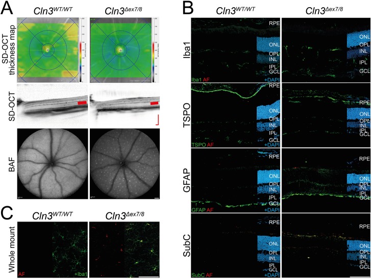
Fig. 1.
Retinal phenotype and gliosis in young CLN3-deficient and wild-type mice. (A) In vivo imaging shows only slight thinning of retinas from 8- to 10-week-old CLN3-deficient mice, indicated by the blueish color in retinal SD-OCT thickness maps. No alteration of retinal structure was observed in CLN3-deficient mice compared to wild-type mouse retinas. BAF imaging shows the occurrence of a constant pattern of autofluorescent spots in the fundus of CLN3-deficient mice and the slightly thinner ONL (red bars in SD-OCT scans). Scale bar: 200 µm. (B) Immunohistochemical stainings of retinal cryosections for the microglia marker Iba1 and the gliosis markers TSPO and GFAP revealed no clear difference between CLN3-deficient and wild-type animals. A consistent staining for SubC was present in CLN3-deficient retinas. Scale bar: 100 µm. (C) Iba1-stained retinal flat mounts of CLN3-deficient mice and wild-type mice show normal morphology of microglia and accumulation of autofluorescent storage material only in microglial somata of CLN3-deficient retinas. Scale bar: 100 µm. BAF, BluePeak blue laser autofluorescence; RPE, retinal pigment epithelium; ONL, outer nuclear layer; OPL, outer plexiform layer; INL, inner nuclear layer; IPL, inner plexiform layer; GCL, ganglion cell layer; AF, autofluorescence; TSPO, translocator protein (18 kDa); GFAP, glial fibrillary acidic protein; SubC, mitochondrial ATPase subunit c.

