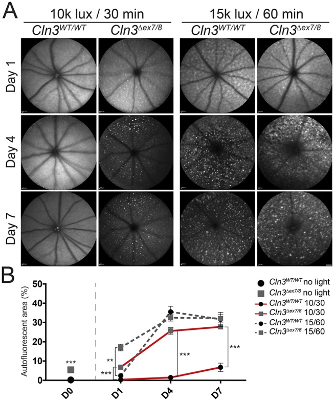
Fig. 3.

Accumulation of autofluorescent material in the fundus of white-light-exposed CLN3-deficient and wild-type mice. (A) BAF imaging shows enriched autofluorescent material in CLN3-deficient retinas after exposure to ‘low light’ conditions, whereas very little autofluorescent material was observed in wild-type animals 7 days after light exposure. Under ‘high light’ conditions, both genotypes show a highly enriched amount of autofluorescent material in the fundus. Scale bar: 1 mm. (B) Comparison of unexposed wild-type and CLN3-deficient retinas shows slight but significant occurrence of autofluorescent material in CLN3-deficient retinas (wild type n=32 eyes, Cln3Δex7/8 n=28 eyes). Quantification of light-exposed data revealed significant difference in autofluorescent storage material of wild-type mice and CLN3-deficient mice under ‘low light’ conditions (n≥15 eyes/time point/group). No differences could be observed between both genotypes after ‘high light’ conditions. No significant difference could be shown between 4 days and 7 days after light exposure (n≥11 eyes/time point/group). Data show mean±s.e.m. from four independent experiments with **P<0.01 and ***P<0.001. The lux value (×1000) and length of exposure (30 or 60 min) is shown in the key. D0, unexposed; D1, 1 day after light exposure; D4, 4 days after light exposure; D7, 7 days after light exposure.
