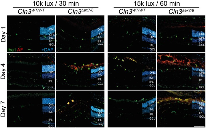
Fig. 4.
Microgliosis in white-light-exposed CLN3-deficient mice. Iba1-positive cells were located in the plexiform layers of wild-type mice exposed to ‘low light’ conditions. Microglial protrusions recognizing the beginning of cell death in the ONL are visible at day 1. Microglial migration towards the subretinal space was visible at 4 and 7 days after light exposure in CLN3-deficient retinas after ‘low light’ and ‘high light’ conditions, and for wild-type retinas only after ‘high light’ conditions. Scale bar: 100 µm. AF, autofluorescence; ONL, outer nuclear layer; OPL, outer plexiform layer; INL, inner nuclear layer; IPL, inner plexiform layer; GCL, ganglion cell layer.

