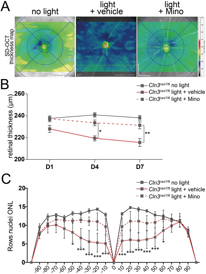
Fig. 6.

Minocycline prevents retinal thinning of CLN3-deficient mice after white light exposure. (A) Representative SD-OCT thickness maps reveal less retinal thinning (green) in minocycline-treated mice compared to vehicle-treated CLN3-deficient mice (blue). (B) Quantification of central retinal thickness shows significant reduction in retinal thinning in minocycline-treated mice after exposure to white light (n≥9 animals/ group). (C) Counting photoreceptor cell nuclei in DAPI-stained cryosections shows the same tendency as the thickness maps (n≥9 eyes/group). The x-axis shows the distance from the optic nerve head in %. Data show mean±s.e.m. from three independent experiments with *P<0.05, **P<0.01, ***P<0.001 light exposed+vehicle versus light exposed+minocycline. D1, 1 day after light exposure; D4, 4 days after light exposure; D7, 7 days after light exposure; Mino, minocycline.
