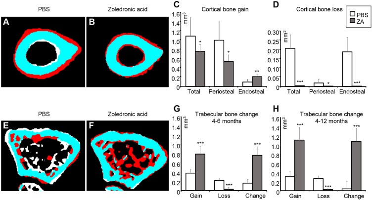
Fig. 6.
Effect of ZA on the bone shape of the p62P394L+/+ mice analysed by μCT. ZA- or PBS-treated p62P394L+/+ mice were scanned using a Skyscan 1076 in vivo µCT scanner (resolution 18 µm) at 4, 6 and 12 months of age. The scans were registered and femoral bone shape changes over time were analysed. (A,B,E,F) White indicates bone lost, red indicates bone gained and blue indicates no change between the time points. A and B show changes at the mid diaphysis between 4 and 12 months of age. PBS-treated p62P394L+/+ mice show endosteal bone loss and periosteal bone gain (A), whereas ZA-treated mice show both endosteal and periosteal bone gain and very little bone loss (B). E and F show changes in the distal femoral metaphysis between 4 and 6 months of age. Note the substantial increase in bone volume in the ZA-treated animal (F). (C,D) Quantification of changes in A and B. (G) Quantification of the trabecular bone volume changes between 4 and 6 months of age. (H) Quantification of the trabecular bone volume changes between 4 and 12 months of age. Data are mean±s.d. *P<0.05, **P<0.01, ***P<0.001 (Student's t-test). PBS, n=6; ZA, n=8. Anchor points added by the CTAn software have been removed from panels A, B, E and F.

