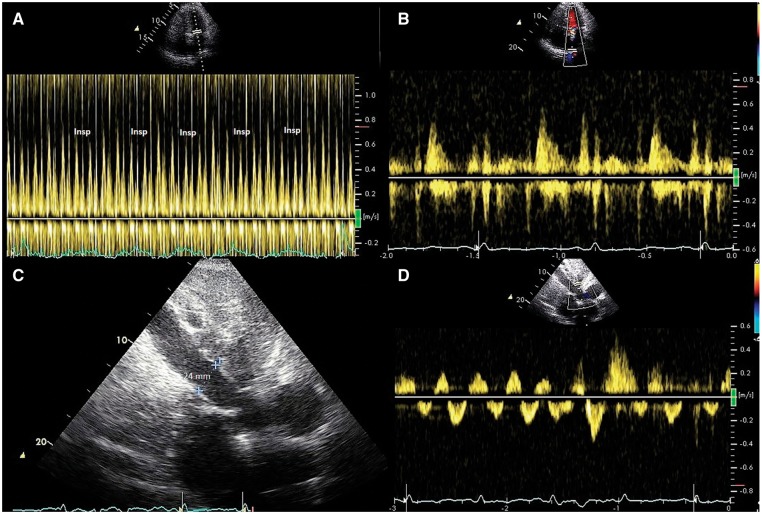
Figure 1.
(A) Pulsed Doppler recording of left ventricular inflow showing reduced early diastolic filling with inspiration. (B) Pulsed Doppler recording of pulmonary vein flow showing a prominent diastolic filling phase. (C) Subcostal view showing a dilated inferior vena cava reflecting the elevated right atrial pressure. (D) Pulsed Doppler recording of hepatic vein flow showing increased hepatic vein flow reversal with expiration.

