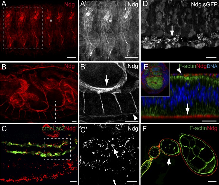
Fig 1. Distribution of the Ndg protein.
(A-F) Confocal images showing embryonic (A-D), wing imaginal disc (E), and ovarian tissues (F) stained with anti-Ndg (A-C, and E-F) or anti-GFP (Ndg.sGFP, D). (A, B) In stage 16 wild-type embryos, Ndg (red) is found in the BMs surrounding most tissues, including muscles (A, A’), gut (B, B’ (arrow) and VNC (B, B’, arrowhead) and in chordotonal organs (asterisk). (C, C’) Lateral view of a stage 13 embryo showing Ndg (red) accumulation around caudal visceral mesodermal cells visualized with the marker crocLacZ (green, arrow in C’). (D) Ndg is found in embryonic macrophages (arrow). (E) Ndg (red) is found at the basal surface of wing imaginal disc epithelial cells (arrow) and cells of the peripodial membrane (arrowhead). (F) Ndg (red) accumulates in the basement membrane (BM) around the follicular epithelium (arrow). Scale bars represent 20μm (A-F).

