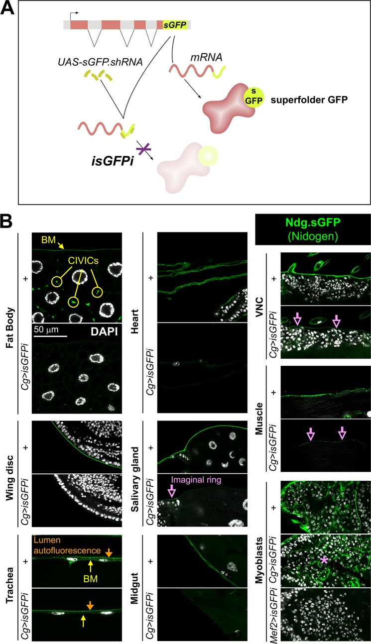
Fig 2. Fat body adipocytes and blood cells are the main source of Ndg in larval BMs.
(A) Schematic representation of the in vivo sGFP interference approach (isGFPi). (B) Confocal images showing the localization of functional Ndg.sGFP fusion protein expressed under control of the endogenous promoter in different tissues of the 3rd instar larva. Images compare tissues from control larvae (+) and larvae where Ndg.sGFP expression has been knocked down in fat body adipose tissue and blood cells (Cg>isGFPi). Ndg.sGFP signal is not observed in the BMs of Cg>isGFPi larvae, except in wing disc myoblasts (asterisk) and partially in the imaginal ring of the salivary gland, body wall muscles and VNC (purple arrows). Note that Ndg.sGFP signal disappears from myoblasts when isGFPi is driven with myoblast driver Mef2-GAL4 (Mef2>isGFPi). Arrow and circles in the fat body panel indicate BM and CIVICs (Collagen IV Intercellular Concentrations), respectively. In tracheal images, apical cuticle autofluorescence is observed in the tracheal lumen (downward-pointing yellow arrow). Nuclei stained with DAPI (white).

