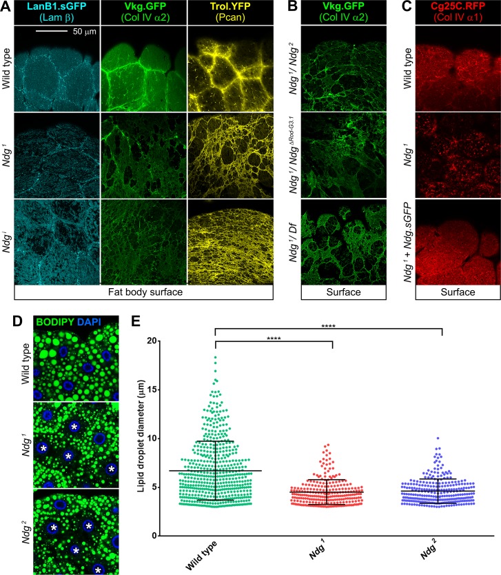
Fig 4. Ndg mutants show discontinuous adipose tissue BMs.
(A) Confocal images of the larvae fat body BM showing localization of Laminin (LanB1.sGFP, cyan), Collagen IV (Vkg.GFP, green) and Perlecan (Trol.YFP, yellow) from control (upper panels), Ndg1 mutant (middle panels) and larvae where Ndg has been knocked down under control of Cg-GAL4 (Ndgi, lower panels). Loss of Ndg causes discontinuity of adipose tissue BMs. (B) Discontinuous BMs (Vkg.GFP, green) in the larval fat body of transheterozygotes Ndg1/Ndg2 (upper panel), Ndg1/NdgΔRod-G3.1 (middle panel) and Ndg1/NdgDf(2R)BSC281 (lower panel). (C) Fat body BM (Cg25C.RFP, red) in wild type, Ndg1 mutant and Ndg1 mutant rescued with Ndg.sGFP. (D) Lipid droplets (neutral lipid dye BODIPY, green) in fat body of wild type (upper panel), Ndg1 mutant (middle panel) and Ndg2 mutant (lower panel) larvae. Asterisks point to cells with reduced content of lipid droplets. Nuclei stained with DAPI (blue). Scale bar represents 50μm (A-D). (E) Quantification of lipid droplet diameter in 16 cells from wild type control, Ndg1 and Ndg2 mutants. Each dot represents a single droplet. Particles smaller than 3μm in diameter were excluded from the analysis. Horizontal lines indicate the mean value and error bars represent ±SD. Difference with the wild type are significant in non-parametric Mann-Whitney tests (****: p<0.0001).

