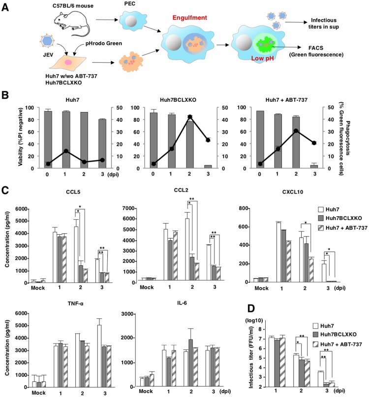
Fig 5. Inhibition of BCLXL suppresses viral production and innate immune responses.
(A) Schematic of in vitro phagocytosis assays. BCLXKO cells and Huh7 cells treated with/without ABT-737 (1 μM), were infected with JEV, labelled with pHrodo green at 1, 2 and 3 days post-infection and overlaid onto PECs pre-incubated overnight. Infectious titers in the supernatants and engulfment by PECs were determined by focus-forming assay and FACS analysis, respectively. (B) The percentages of engulfed cells were determined as green fluorescence by FACS analysis (line, right axis). Viability of JEV-infected BCLXKO cells and JEV-infected Huh7 cells treated with/without ABT-737 (1 μM) was assessed at indicated time points (bar, left axis). The data represent the mean ± SD of two independent experiments. (C) Suppression of viral propagation by phagocytosis of infected cells. JEV-infected BCLXKO cells and JEV-infected Huh7 cells treated with/without ABT-737 (1 μM) were overlaid onto PECs at 1, 2 and 3 days post-infection. Infectious titers in the culture supernatants were determined by focus-forming assay at 1 day after incubation. The data represent the mean ± SD of three independent experiments performed with a culture representative of each cell line. (D) Production of TNF-α, CCL5, CCL2, IL-6 and CXCL10 in the supernatants of PEC cultures (generated as in Figure 5C) was determined by ELISA. The data are representative of three independent experiments. Significant differences (C, D) were determined using Student’s t-test and are indicated with asterisks (*P<0.05) and double asterisks (**P<0.01).

