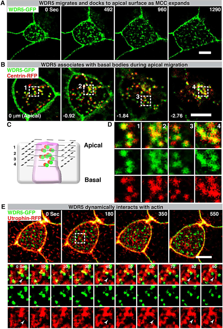
Figure 6: WDR5-basal body complex interacts with F-actin during apical expansion.
(A) A montage of Xenopus epidermal MCC undergoing apical expansion over 21 minutes. WDR5 marked with WDR5-GFP localizes to the apical membrane as MCC expands.
(B,D) A montage of Xenopus epidermal MCC expressing WDR5-GFP and centrin-RFP early during MCC expansion process. Basal bodies (centrin-RFP) and WDR5 appear to co-localize deep in the cytoplasm.
(C) Schematic showing optical sections of a MCC to examine WDR5 and basal body colocalization in B and D. Optical section 1–4 correspond to the optical sections in the panels B and D.
Scale bar = 5 μM
(E) A montage of Xenopus epidermal MCC undergoing apical expansion over 9 minutes. WDR5 (WDR5-GFP) interacts with F-actin (Utrophin-RFP) at the apical surface as MCC expands. The region in a white square is magnified to show F-actin organizing around WDR5 (note white arrowheads).

