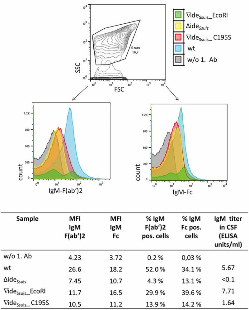
Figure 11.

S. suis in the CSF of experimentally infected piglets is labeled with porcine IgM. Eight-week old piglets were intranasally infected with S. suis strain 10 (wt), 10∆ideSsuis (∆ideSsuis), 10∆ideSsuis∇ideSsuis_EcoRI (∇ideSsuis_EcoRI) and 10∆ideSsuis∇ideSsuis_C195S (∇ideSsuis_C195S), respectively. The CSF of one pig with meningitis of each infection group was centrifuged and the intracerebrospinal S. suis population stained with anti-IgM F(ab’)2 and anti-IgM Fc specific primary antibodies and phycoerythrin (PE) and fluorescein (FITC) labeled secondary antibodies. Bacteria were analyzed for the presence of IgM on their surface via flow cytometry. S. suis stained without the first antibodies served as negative control (w/o 1. Ab). The upper panel shows the gating strategy used to define the S. suis population. The lower panels shows the S. suis population in the CSF of infected piglets as overlay histograms of the F(ab’)2 signals (left hand side) and the Fc signals (right hand side). The table beneath the figure shows the geometric mean fluorescence intensity (MFI), the percentage of IgM F(ab’)2 and IgM Fc positive bacteria and the IgM titers of the four representative piglets.
