. 2018 Sep 21;7:e35316. doi: 10.7554/eLife.35316

Figure 5. Hypoxia acts via mitochondrial superoxide to induce endothelium-dependent dilation of pressurized cerebral arteries.

(A) Representative traces (left) and summary data (right) showing hypoxia-induced dilation in intact and endothelium-denuded cerebral pial arteries (*p<0.05, Student’s t-test; n = 5 arteries from three different mice). (B) Representative traces of lumen diameter of a pressurized cerebral pial artery (left) and summary data (right) showing hypoxia-induced dilation in the presence of the cell-permeant SOD mimetic Tempol (100 µM). (*p<0.05, Student’s t-test; n = 5 arteries from three different mice.) (C) Representative traces of the lumen diameter of a pressurized cerebral pial artery (left) and summary data (right) showing hypoxia-induced dilation in the presence of the cell-permeant mitochondrial membrane-targeted SOD mimetic mitoTEMPO (500 nM). (*p<0.05, Student’s t-test; n = 6 arteries from three different mice). The arteries used for pressure myography experiments were not treated with EGTA-AM or CPA.

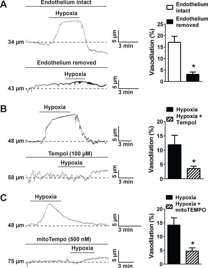
Figure 5—source data 1. Excel spreadsheet containing the individual numeric values of the parameters analyzed in Figure 5.
DOI: 10.7554/eLife.35316.039

Figure 5.

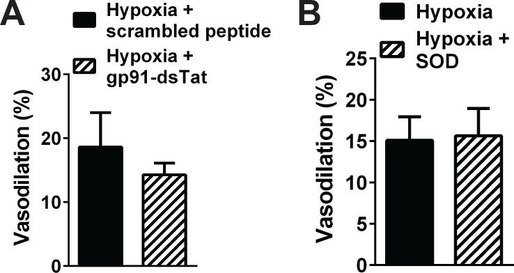
Figure 5—figure supplement 1. NOX2 inhibition and quenching of extracellular O2-and H2O2 did not significantly alter cerebral artery dilation induced by hypoxia.

Figure 5—figure supplement 1.

Summary data showing that hypoxia-induced dilation of cerebral pial arteries was not significantly altered by NOX2 inhibition with gp91-dsTat (1 μM, (A), and by removal of extracellular O2 with SOD (200 U/mL, (B). n = 3 arteries from three different mice for the gp91-dsTat experiments, and n = 5 arteries from three different mice for the extracellular SOD experiments. Arteries used for pressure myography experiments were not incubated with EGTA-AM or CPA.
Figure 5—figure supplement 1—source data 1. Excel spreadsheet containing the individual numeric values of the parameters analyzed in Figure 5—figure supplement 1.
DOI: 10.7554/eLife.35316.036
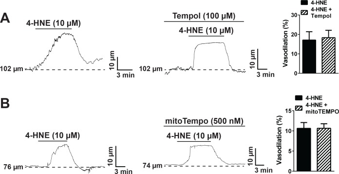
Figure 5—figure supplement 2. Superoxide dismutase mimetics do not directly inhibit TRPA1 channels.

Figure 5—figure supplement 2.

(A) The cell permeable superoxide dismutase (SOD) mimetic Tempol (100 µM) did not alter dilation of pressurized cerebral arteries to the TRPA1 agonist 4-hydroxynonenal (4-HNE), as observed by the representative traces on the left and middle, and summary graph on the right. N = 3 arteries from three different mice. (B) Similarly, the mitochondria-targeted SOD mimetic mitoTEMPO (500 nM) did not affect TRPA1-mediated dilation caused by exposure to 4-HNE. N = 5 arteries from three different mice. Arteries used for pressure myography experiments were not incubated with EGTA-AM or CPA.
Figure 5—figure supplement 2—source data 1. Excel spreadsheet containing the individual numeric values of the parameters analyzed in Figure 5—figure supplement 2.
DOI: 10.7554/eLife.35316.038