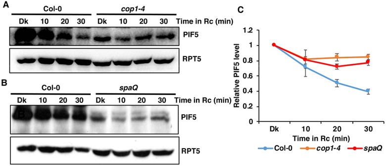
Figure 1. Red light induced PIF5 degradation is slower in cop1–4 and spaQ mutants.
(A-B) Immunoblots showing the degradation pattern of endogenous PIF5 in response to dark and constant Rc in cop1–4 (A) and spaQ (B) mutants, compared to the wild-type seedlings. Four-day-old dark-grown seedlings were treated with either dark (Dk) or 2 μmolm−2 s−1 constant red light (Rc) for the indicated period of time (min). Total protein was extracted and resolved on 8% SDS-PAGE gel. Proteins were transferred to PVDF membrane and sequentially probed with anti-PIF5 and anti-RPT5.
(C) Line-graph shows the relative rate of degradation of endogenous PIF5 in response to constant Rc over the dark treatment. Band intensities of PIF5 and RPT5 from three replicates were measured using ImageJ tool. For each of the genotype, PIF5 level in the dark (Dk) was set to 1and the relative PIF5 level in response to constant Rc was calculated.

