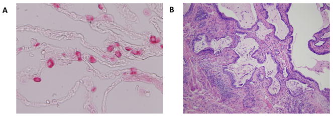
Fig. 1. The pulmonary ECM in health and disease.
Image A depicts the normal alveolar space at high power. The type 2 pneumocytes are stained pink. There is minimal matrix present and the alveolar epithelium is very thin allowing for ease of gas exchange. Image B depicts fibroblastic foci of IPF, with layering down of aberrant ECM and the loss of the normal alveolar epithelium leading to impaired gas exchange.

