
Figure 2. High-resolution structures of hnRNP A1 complexes with DNA and RNA.
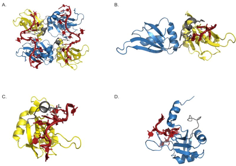
(A) The crystal structure of the UP1-telomeric DNA complex (2UP1). The complex crystalized as a homodimer with a 2:2 stoichiometry. (B) The crystal structure of the UP1-rAGU complex (4YOE). The structure crystalized as a monomer with 1:1 stoichiometry where the 5′-rAGU-3′ trinucleotide was shown to interact only with the RRM1 surface and inter-RRM linker. The NMR solution structures of the (C) RRM1-RNA (5MPG) and (D) RRM2-RNA complexes (5MPL). For each structure, select amino acid side chains that form the binding pockets on the surface of RRM1 and RRM2 of each are shown as ball and sticks. The core rAG dinucleotides that are specifically recognized by hnRNP A1 are also labeled.