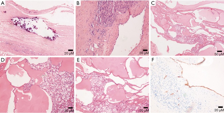
Figure 15.
(A-E) Photomicrographs (original magnification, A,B: ×100; C: ×40; D,E: ×100; H-E stain) show calcification in (A) lymphangiomas lined by flattened endothelial cells with no significant atypia in (B) and lymphatic fluid and dilated lymphatic ducts are found in (C-E). (F) Photomicrograph (original magnification, ×100; D2-40 IHC stain) presents positive result.

