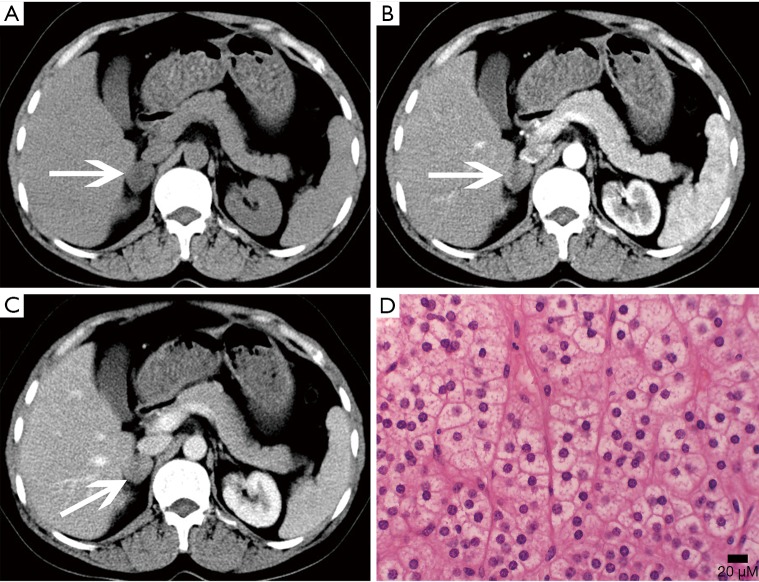
Figure 2.
Adrenocortical adenoma confirmed with pathology in a 43-year-old woman who presented with right adrenal mass for 4 years. (A) Axial pre-contrast CT image shows a 21 mm × 20 mm mass with clear margin and heterogeneous density (arrows); (B) axial arterial and (C) venous phase images show moderate enhancement; (D) photomicrograph (original magnification, ×400; H-E stain) shows the tumor cells are similar to normal cortical cells, with pale-staining cytoplasm.

