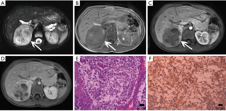
Figure 28.
Neuroblastoma confirmed with pathology in 4-year-old boy. (A) Axial T2-weighted image, (B) axial T1-weighted image show a 62 mm × 54 mm oval mass with mixed signal (arrows). (C-D) Enhanced MR imaging demonstrates marked heterogeneous enhancement. (E) Photomicrograph (original magnification, ×400; H-E stain) shows the tumor consists of ganglion cells and ensheathing cells. (F) Photomicrograph (original magnification, ×200; immunohistochemical staining): NSE (+).

