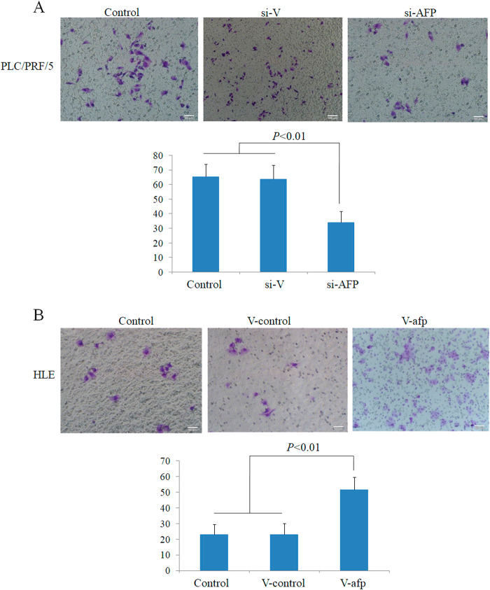
Fig. 7. Effects of AFP on the invasion of PLC/PRF/5 and HLE human liver cancer cells.
a PLC/PRF/5 cells were transfected with AFP-siRNA vectors for 48 h, and invasive cells were stained with 0.1% crystal violet and observed by microscopy; the lower column graph indicates the quantity of invasive cells. P < 0.01 versus control and scrambled-siRNA groups. b HLE cells were transfected with pcDNA3.1-afp for 48 h, and invasive cells were stained with 0.1% crystal violet and observed by microscopy; the lower column graph indicates the quantity of invasive cells. P < 0.01 versus control and pcDNA3.1-vector groups. Three independent experiments were performed to generate these data

