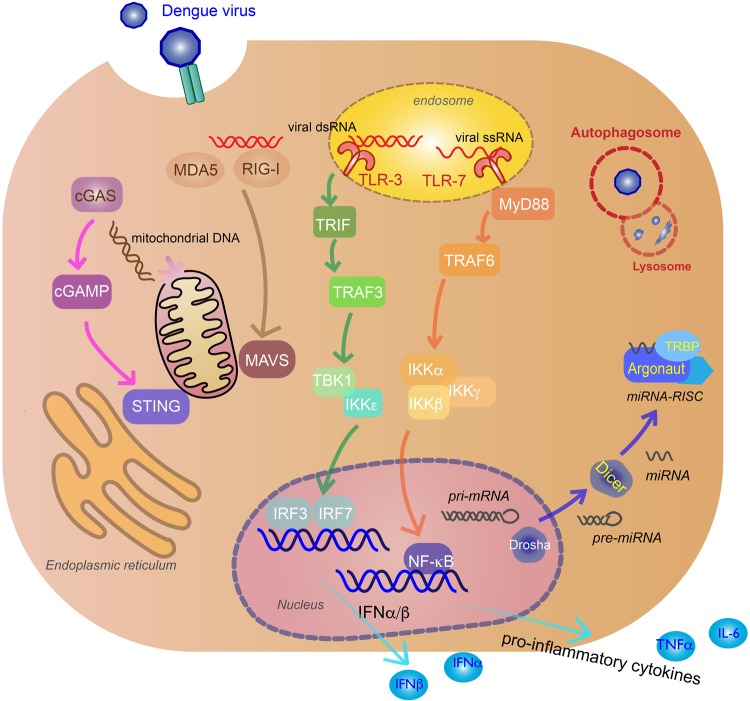
Fig. 2. Innate immune response to DENV infection.
Recognition of viral genomes by cytoplasmic retinoic acid-inducible gene I (RIG-I) and melanoma differentiation-associated protein 5 (MDA5) trigger mitochondrial antiviral signaling (MAVS) activation that lead to TANK-binding kinase 1 (TBK1), IκB kinase-ε (IKKε) induction of interferon regulatory factor 3 (IRF3), and IRF7 (tan arrows). Viral genome recognition by endosomal toll-like receptor 3 (TLR3) (green arrows) and TLR7 (orange arrows) will activate TIR-domain-containing adapter-inducing IFNβ (TRIF) and myeloid differentiation primary response gene 88 (MyD88) signaling pathways, inducing IRF3/IRF7 and inhibitor of nuclear factor-κB kinase (IKK)α/IKKβ/IKKγ, and activate nuclear factor-κB (NF-κB) to produce IFNα/β and pro-inflammatory cytokines. Virus-induced mitochondria damage activates the cyclic GMP-AMP synthase (cGAS) (pink arrows) and stimulator of interferon gene (STING) pathway to induce IFNα/β production via IRF3 and IRF7. MicroRNAs (miRNAs) associate with RNA-induced silencing complex (RISC) in the cytoplasm to target viral RNA for inhibition or degradation. miRNA biogenesis starts in the nucleus as pri-miRNA and processed by Drosha into pre-miRNA. Pre-miRNAs are transported to the cytoplasm and cleaved by Dicer to produce mature miRNAs100. Argonaute (Argo) and TAR RNA-binding protein (TRBP) are proteins essential to the formation of RISC (blue arrows). Double membrane vacuoles, called autophagosomes, will engulf foreign cytoplasmic material and fuse with the lysosome for degradation, inhibiting virus replication (red)

