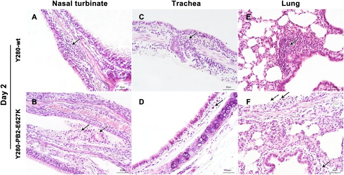
Fig. 3. Histopathology of the respiratory epithelium at 2 dpi in tree shrews infected with H9N2 viruses.
Tree shrews (n = 4 per group) were infected with 106 TCID50 of one of two H9N2 viruses (Y280-wt or Y280-PB2-E627K). Nasal turbinate (a, b), trachea (c, d), and lung (e, f) tissues were collected at 2 dpi, processed into paraffin sections and stained with H&E. Black arrows indicate infiltration of inflammatory cells, and dotted arrows indicate necrotic and sloughed epithelial cells. The images are shown at ×400 magnification

